PUBLICATIONS

Breast Tumor Malignancy Prediction in Ultrasound

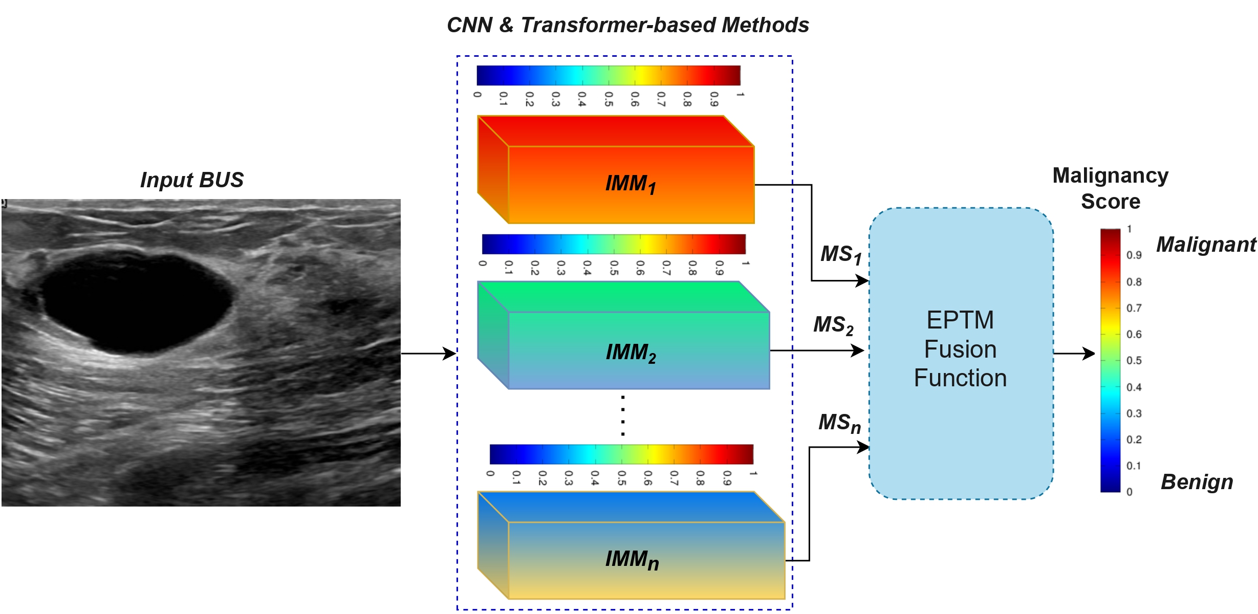
This work proposes an efficient method for predicting breast tumor malignancy (EPTM) in BUS images. EPTM comprises heterogeneous deep learning-based feature extraction models and a Choquet integral-based fusion mechanism. Specifically, EPTM incorporates efficient CNN and vision-Transformer methods to build accurate individual breast tumor malignancy prediction models (IMMs).

Vivek Kumar Singh, Ehab Mahmoud Mohamed, and Mohamed Abdel-Nasser. ” Aggregating efficient transformer and CNN networks using learnable fuzzy measure for breast tumor malignancy prediction in ultrasound images.” Neural Computing and Applications (2024): 1-17.
True-T: Improving T-cell Response Quantification

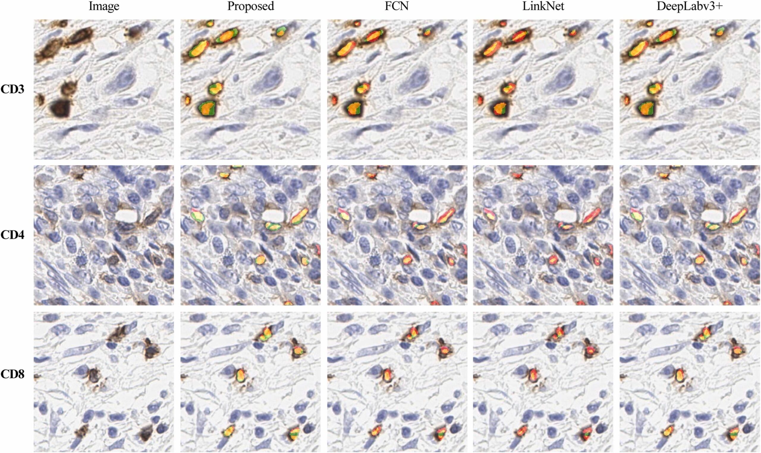
We present a new method called True-T, which employs artificial intelligence-based techniques to quantify T-cells in colorectal cancer (CRC) using IHC images. True-T analyses the chromogenic tissue hybridization signal of three widely recognized T-cell markers (CD3, CD4, and CD8). Our method employs a pipeline consisting of three stages: T-cell segmentation, density estimation from the segmented mask, and prediction of individual five-year survival rates.

Makhlouf, Yasmine, Vivek Kumar Singh, Stephanie Craig, Aoife McArdle, Dominique French, Maurice B. Loughrey, Nicola Oliver et al.” True-T–Improving T-cell response quantification with holistic artificial intelligence-based prediction in immunohistochemistry images.” Computational and Structural Biotechnology Journal 23 (2024): 174-185.
Detecting Breast Tumors in Tomosynthesis Images

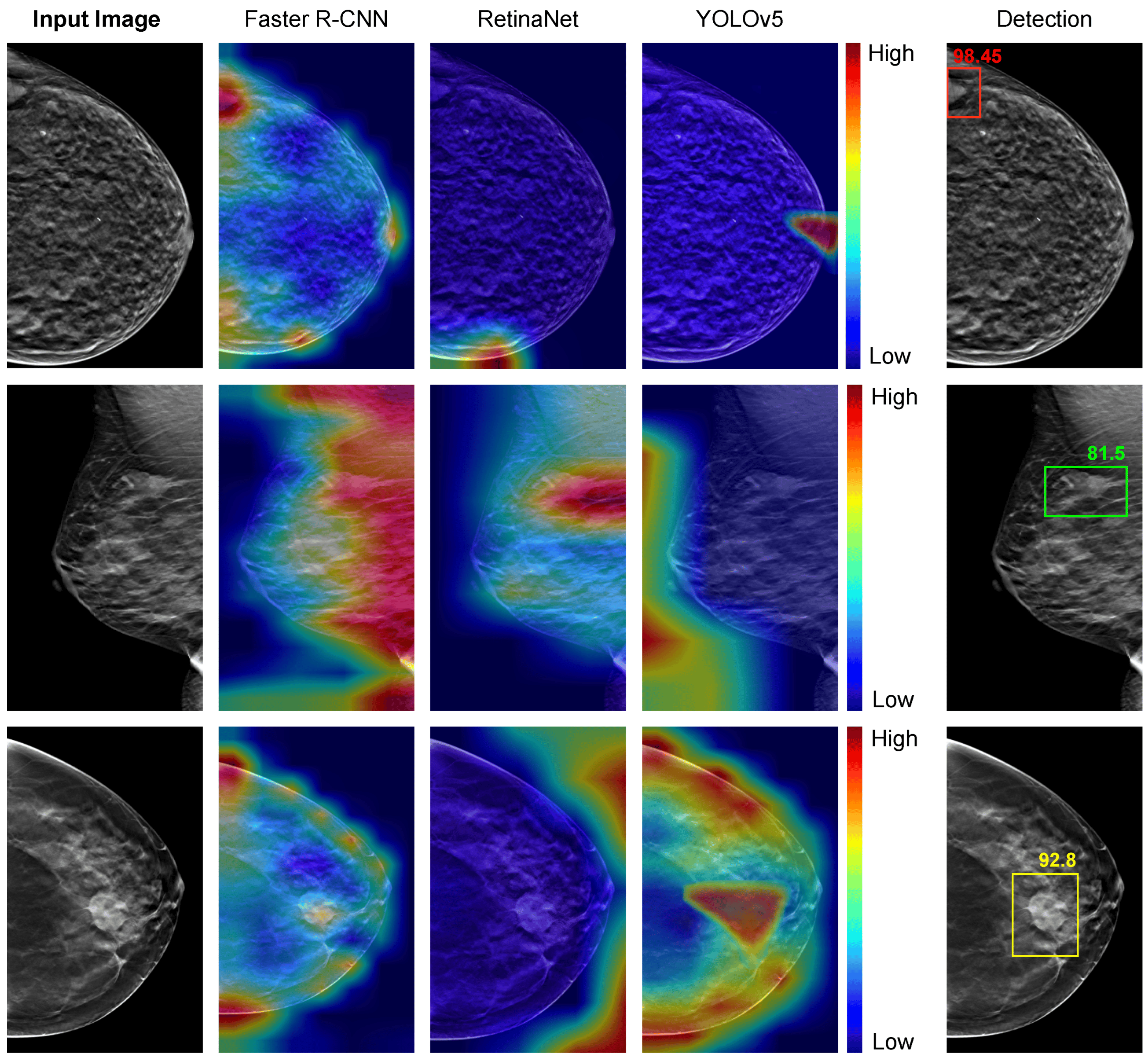
We propose a novel method for detecting breast tumors within Digital breast tomosynthesis (DBT) images. This method relies on a potent dynamic ensemble technique, along with robust individual breast tumor detectors (IBTDs). The proposed dynamic ensemble technique utilizes a deep neural network to select the optimal IBTD for detecting breast tumors, based on the characteristics of the input DBT image.

Hassan, Loay, Adel Saleh, Vivek Kumar Singh, Domenec Puig, and Mohamed Abdel-Nasser. ”Detecting Breast Tumors in Tomosynthesis Images Utilizing Deep Learning-Based Dynamic Ensemble Approach.” Computers 12, no. 11: 220.
COMFormer: Classification of Maternal–Fetal and Brain Anatomy

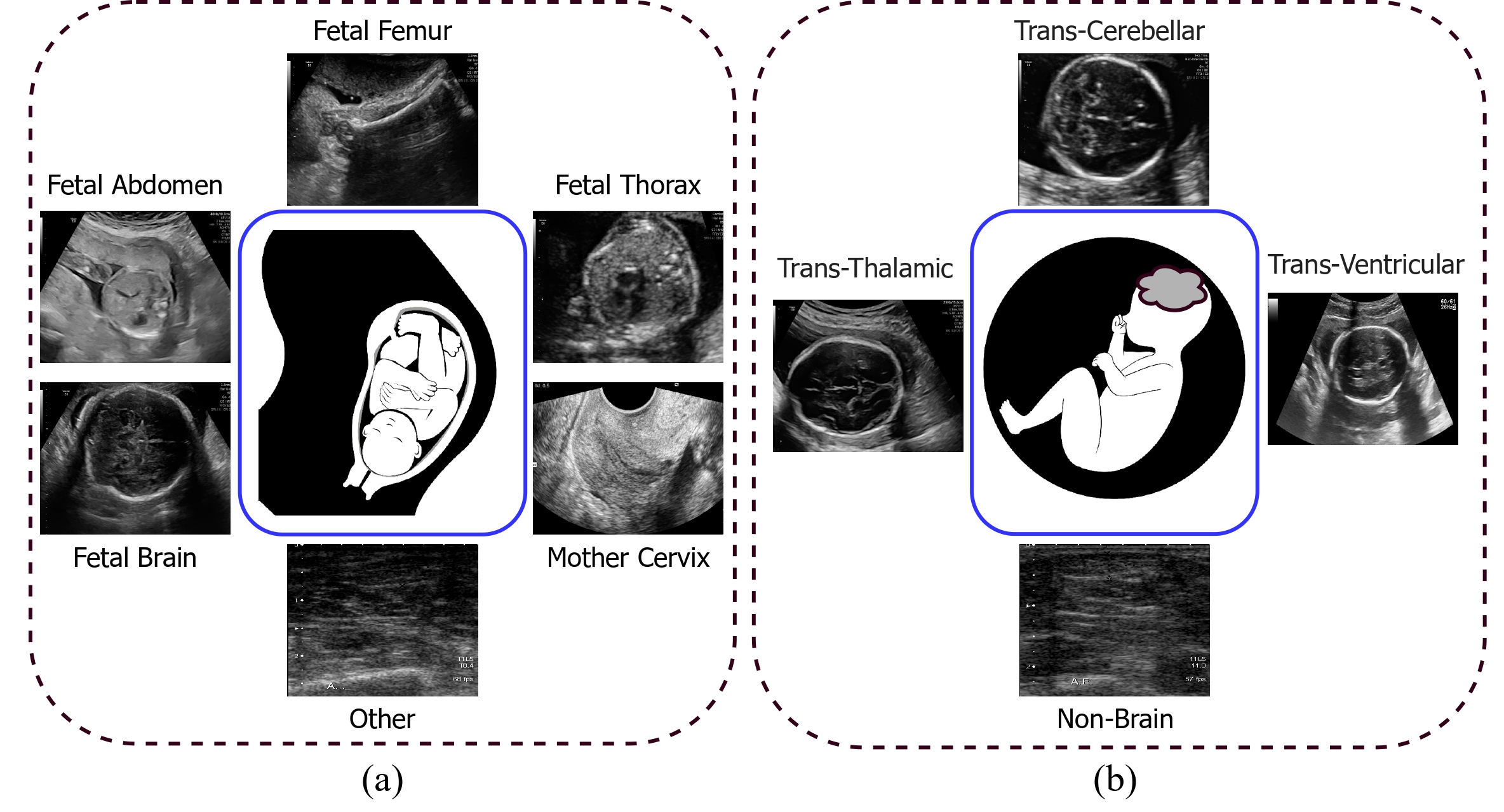
We propose a deep-learning-based image classification architecture called the COMFormer to classify maternal-fetal and brain anatomical structures present in 2-D fetal ultrasound (US) images. The proposed architecture classifies the two subcategories separately: maternal-fetal (abdomen, brain, femur, thorax, mother’s cervix (MC), and others) and brain anatomical structures [trans-thalamic (TT), trans-cerebellum (TC), trans-ventricular (TV), and non-brain (NB)]. Our proposed architecture relies on a transformer-based approach.
Sarker, Md Mostafa Kamal, Vivek Kumar Singh, Mohammad Alsharid, Netzahualcoyotl Hernandez-Cruz, Aris T. Papageorghiou, and J. Alison Noble. “COMFormer: classification of maternal-fetal and brain anatomy using a residual cross-covariance attention guided transformer in ultrasound.” IEEE Transactions on Ultrasonics, Ferroelectrics, and Frequency Control (2023).

Current and Emerging Trends in Medical Image Segmentation

We examine the numerous developments and breakthroughs brought since the rise of U-Net-inspired architectures. Especially, we focus on the technical challenges and emerging trends that the community is now focusing on, including conditional generative adversarial and cascaded networks, medical Transformers, contrastive learning, knowledge distillation, active learning, prior knowledge embedding, cross-modality learning, multistructure analysis, federated learning.
Conze, Pierre-Henri, Gustavo Andrade-Miranda, Vivek Kumar Singh, Vincent Jaouen, and Dimitris Visvikis. “Current and emerging trends in medical image segmentation with deep learning.” IEEE Transactions on Radiation and Plasma Medical Sciences (2023).

Staining-Invariant Nuclei Segmentation using Self-Supervised Learning

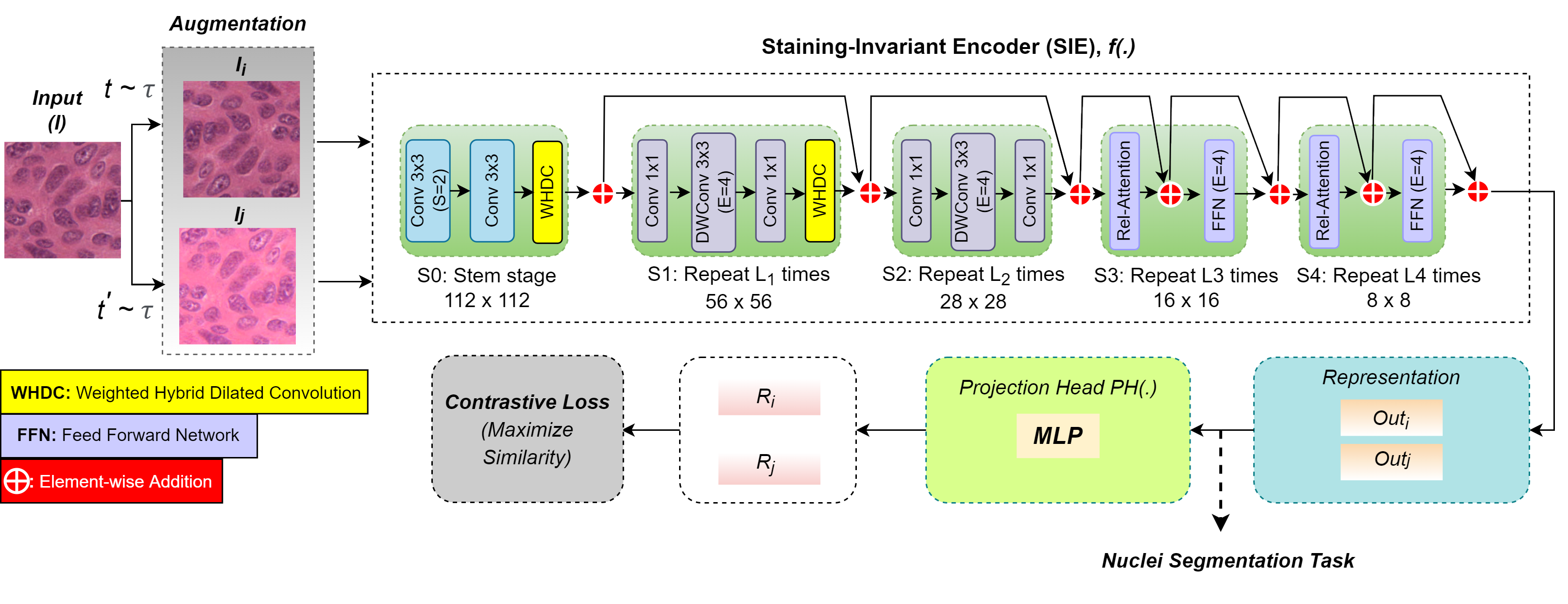
We proposes an efficient staining-invariant nuclei segmentation method based on self-supervised contrastive learning and an effective weighted hybrid dilated convolution (WHDC) block. In particular, we propose a staining-invariant encoder (SIE) that includes convolution and transformers blocks. We also propose the WHDC block allowing the network to learn multi-scale nuclei-relevant features to handle the variation in the sizes and shapes of nuclei.
Abdel-Nasser, Mohamed, Vivek Kumar Singh, and Ehab Mahmoud Mohamed. “Efficient Staining-Invariant Nuclei Segmentation Approach Using Self-Supervised Deep Contrastive Network.” Diagnostics 12, no. 12 (2022): 3024.
© Copyright AI4MEDTECH. All Rights Reserved.